About hyaluronic acid
Dog joint health
Recognizing the signs:

- Are they slow getting up from a resting position?
- Do they have trouble getting in the car or up the stairs?
- Are they much slower on recent walks?
- Are they sensitive to touch and become aggressive?
It is important to observe your dog closely:
- Decreased activity?
- Reluctance to walk, run, jump or play?
- Lagging behind on walks?
- Difficulty rising from a resting position?
- Acting aggressive or withdrawn?
- Exhibiting other personality changes?
If you notice any of these changes, consult your veterinarian. The sooner the condition is recognized, the sooner your dog can be active again!
Understanding joint anatomy:

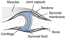
To understand joint health, one must become familiar with the anatomy of a healthy functioning joint. Let’s start off with understanding the joint capsule. The joint capsule is a thick fibrous tissue that connects the bones, provides the outer layer of the joint and holds the fluid inside the joint cavity. The muscles serve to support the joint capsule and to provide joint movement. The tendon is an elastic cord that attaches the muscle to the bone and assist with movement. The synovial membrane is the inner lining of the joint capsule. It is highly vascularized and therefore is responsible for carrying nutrients to the joint and most importantly, producing the synovial fluid. The synovial fluid is a clear viscous fluid that lubricates the joints. It consists of blood plasma and hyaluronic acid. Without it, joint movement would be limited and articular cartilage becomes vulberable. The cartilage covers the ends of the bones and absorbs most of compression and stress in the joint. Because it is a slippery material, it allows the joints to move smoothly and easily.
Overall, the parts of the joint have to work together, but the two most important parts are the synovial fluid which provides the lubrication for the joint and the cartilage, which absorbs the stress. When these two are damaged, problems are unavoidable.
How veterinarians recognize problems:

- First your vet will evaluate the case history of your dog. As part of this backgrounding your vet may ask you several questions about your dog’s care and activities to determine a potential cause of the joint concern.
- Next your vet will do a physical examination of your dog. This usually involves palpating the areas of concern. Most vets are very skilled with their hands and can often feel if a joint is not functioning correctly.
- The veterinarian will inspect your dog at a walk and a run. They will walk them in a straight line watching for any abnormalities.
- Your veterinarian may perform a flexion test. This is done by bending the joint and holding for at least one minute and then released. After the joint is released, the dog is observed while walking and running. Often times this will exaggerate the condition and make it more clearly visible during activity.
- It may be necessary to perform a radiographic x-ray examination to get a visual representation of what might be the cause.
- Your veterinarian may remove by needle some of the synovial fluid located in the joint to determine if an infection is present.
Deterring health problems:
Older dogs have a variety of health problems due to a number of environmental stressors, but you can help relieve some of these stresses by:
- Avoiding obesity and heavy loads
- Providing your dog with suitable bedding
- Avoiding quick changes in duration or intensity of exercise
- Avoiding hard and unstable ground surfaces
- Feeding a diet high in protein and other nutrients. Joints can never repair or become stronger without proper nutrition
In summary:
Dog owners should become very familiar with recognizing joint health in their pets. Because joint problems are progressive, acting early can give your dog a better chance at getting back to their normal activities such as walking and running. Often times, the joint is far past repair and is quite likely that the animal will never regain normal movement.
Ligaments, tendons, and connective tissue

Connective tissue is found everywhere in the body. It does much more than connect body parts; it has many forms and functions. Its major functions include binding, support, protection, and insulation. One such example of connective tissue is the cordlike structures that connect muscle to bone (tendons) and bone to bone (ligaments). In all connective tissue there are three structural elements. They are ground substance (hyaluronic acid), stretchy fibers (collagen and elastin) and a fundamental cell type. Whereas all other primary tissues in the body are composed mainly of living cells, connective tissues are composed largely of a nonliving ground substance, the hyaluronic acid, which separates and cushions the living cells of the connective tissue. The separation and cushioning allow the tissue to bear weight, withstand great tension and endure abuses that no other body tissue could. All of this is made possible because of the presence of the HA and its ability to form the gelatinous ground substance fluid.
Skin and scalp tissue

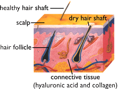
The hair follicle that produces that fur that makes your dog so soft, is actually a derivative of skin tissue. There are two distinctive skin layers, one, the epidermis (outer layer) which gives rise to the protective shield of the body and the other, the dermal layer (deep layer) which makes up the bulk of the skin and is where the hair follicle is located. This dermal layer is composed of connective tissue and the connective tissue, with its gelatinous fluid like characteristics provides support, nourishes and hydrates the deep layers of the scalp.
Again, all of this is made possible because of the presence of HA in the scalp tissue and its ability to form this fluid and hold water.
For more information on hyaluronic acid...
If you would like more information on hyaluronic acid and its uses, please visit the Clinical Research with Hyaluronic Acid page on the Hyalogic corporate website.